|
A.
Plants Animals: 1.
This opposition between autotrophic
and heterotrophic cells concerns
the chemical level, appearing already
among the prokaryotic unicellular
groups.(These include autotrophic
species that lived on energy from
small molecules of non-metals as
sulfur. Disregarded here.)
Plants
have both the photosynthesis with
primary synthesis of carbohydrates
and the respiration cycle, animals
only the latter. Its a relation
of double direction versus one-way
direction, a polarization, one principal
aspect on steps towards lower degrees
in the dimension model.
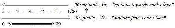
2.
The essential polarity of directions
from 0 and 00-poles appear in the
development of plants and animals:
-
the growth outwards as from a 0-pole
of stem and roots upwards and
downwards - from the seed in plants,
- the growth inwards from a
00-pole, from anticenter of the
blastula, vegetative and animal
poles in animals:
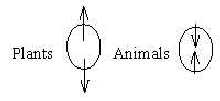 Fig
Ev-add-1-166 Fig
Ev-add-1-166 Plants: divergence
outwards. Animals: convergence inwards. Cf.
that the upward transportation in
plants doesnt demand energy, while
the downward transportation of synthesized
material does. The
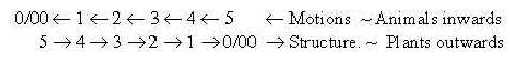
directions of embryonic growth can
be related to definitions in last
step of the dimension chain, motions
from and to each other:
Fig
Ev-add-2-193
3.
A dimension d-degree (d-degree)
step 4 to
3: plants; ↑ ↓, animals → .
In
coordinate axes plants represent
the vertical axis, while later classes
of animals come to represent the
horizontal one, parallel to surface
of the ground: an angle step 180°
→
90° as assumed in
d-degree step 4 →
3; it may also be seen as a difference
radial versus circular, poles 3b
and 3a in our model, corresponding
to the growth of vegetative
pole versus animal pole of
the animal
embryo. These geometries
could be compared with unicellular
algae (autotrophic cells) versus
bacteria: - most algae grow
in long unbranched or branched threads,
-
bacteria more often solitary, round
or staff- or spiral-shaped. Hence,
the growth of algae may reflect
vector fields as d-degree 4, while
next step to 3 implies individualization
(~ rooms, volumes) to closed, separate
forms, in step 3 →
2 transformed to tube forms and
spirals (as from a 3-dimensional
motion).
As
expression for a more high dimensional
field level in plants we can also
see their special type of symplasmatic
cell contacts with cells that
share cytoplasm via long projections
without demarcating, separating
walls (Kz p. 150).
4. Further, the
communication in multicellular plants
is chemical, based on molecules,
while multicellular animals develop
the electrical, ion-based system
as well: a d-degree step associated
with mass versus charge. In the
dimension model here we have proposed
the FA-FG-forces
developed in step 4– 3, the electromagnetic
force (FEM)
in step 3 –
2.
One could
ask if the difference in inner chemistry
is the primary factor for the autotrophic
plant cell - or possibly a secondary
result of cell positions
in relation to a water surface and
sunlight, which have become "anchored"
in the genes? Cf. that chlorophyll
needs sunlight for fulfillment of
its structure and the other metabolic
way from porphyrins goes to animal
hemoglobin.
5.
Another elementary difference Plants
Animals is the one in mobility.
Most outer motions of plants are
bound to their growth, besides adaptations
to light. In animals the growing
number of motional moments from
polarization steps according to
the dimension model gets trapped
within them, become built-in within
their "shells".
The
opposition may be associated with
the polarity of directions: a dimension
chain outwards as structures corresponds
to an opposite chain inwards of
motions ("debranched degrees"
as external motions).
 Fig
Ev-add-3 Motions of plants
is just expressed in their structural
growth, in animals in their external
mobility.
6. There
are also similar structures built
out and built-in respectively. The
tree-like structure up- and down,
turned outside-in in our lungs is
one example, with their branches
into alveoli. The backbone with
the neural tube as a stem and brain
as crown of a tree could be regarded
as another example with the essential
difference that a human brain with
cortex also has circular networks:
cf. circular geometry (pole 3a)
from 00-pole, radial (pole 3b) from
0-pole in the dimension model.
(Crown
of a tree is a receiver of light,
brain a receiver of light and other
sensory impulses.)
7.
The bases for classifications among
animals have parallels among plants
even if not used in the same way
in primary systematics:
- Naturally
1- to multicellular organisms.
-
Number of tissue layers has similarities
with plants with 1 versus 2 cotyledons
and with the division herbs trees:
plants having only the primary growth
of thickness, trees with also the
secondary one and cambium as a kind
of "mesoderm".
- The division
between "thallophytes" and
vascular plants corresponds
most closely to the division between
sponges or 12-layer animals versus
3-layer animals with differentiation
of organs.
- The oppositions
in directions between Proto- and
Deuterostomia have obvious parallels
in plants where seeds grow attached
to either a central structure or
peripherally in the ovary, or stamens
developing either from inside or
from outside. Such differences are
given a systematic value in botany.
One rather curious
thing, similar among plants and
animals, is how their "extremities"
develop, not from the center but
with origin further out, just "under
the skin": branches of trees with
their germs up in the trunk and
"arms legs" of animals built from
near the surface, which also by
the way is the case for "extremities"
as cilia. Connections inwards primary
centers seem secondary. (Fens and
extremities for locomotion and/or
catching of food could be compared
with branches with leaves for catching
of light and CO2).
Is
there anything that really proves
that development is quantified in
dimensional steps it should be such
facts?
(In
the dimension model its a step
from the surface, d-degree 2, to
"linear" structure, d-degree 1:
in the loop version connected with
forces of d-degree 4, outwards -inwards.)
8.
A direct parallel in evolution of
plants and animals is of course
the development toward more and
more built-in and sheltered embryos:
from spread of spores to gymnosperms
to angiosperms in plants, from egg-laying
species to mammals among animals.
B. Prokaryotic
Eukaryotic cells: This
division concerns the cell level,
cells naturally here regarded as
5-dimensional units. Theories about
the emergence of eukaryotic cells
are dealt with in the end of file
The
cell, No. 11. Here
some other annotations. There
are several parallels between this
opposition and the features for
classification among multicellular
organisms: - 1-cellular versus
multicellular organisms: Colonies
of cells exist in both prokaryotic
cells (PKc) - and eukaryotic cells
(EKc), hence a kind of cell contacts,
(in the level
chain corresponding to step
1 ← 0/00),
but PKc remain unicellular while
EKc type leads further to multicellular
organisms. - 1-, 2-, 3-layer
animals: This division corresponds
to number of membranes on the tissue
level, only one (with certain reservations,
Bc p. 290) in Prokaryotic
cells, 2 to 3 in EKc with nuclear
membrane. The endoplasmic reticulum
appear as analogous to the 3rd
layer (mesoderm) in 3-layer organisms.
(Its also a parallel to blastula
versus gastrula,) - Coelom
and body cavity differentiation:
The several organelles within membranes
within EKc are naturally a parallel
to specialized and centralized organs
on the multicellular level a 3-dimensional
development in the interpretations
here.
However,
in contrast to coelom there don't
seem to exist any partial steps
in the development of organelles
among EKc (?), as if it immediately
followed in d-degree step 3 - 2.
- Protostomia Deuterostomia:
at least one certain feature appears
as a parallel on the unicellular
level: the observation that the
cell walls at cell division is created
from outside inwards in PKc, from
inside outwards in EKc (Fb p.
31).
Other
such oppositions are dealt with
in file Centrioles
- Cilia: the central but
hollow tube of PKc flagella, the
center - anticenter organization
of cilia in cilia of EKc; transportation
of building material in PKc inside
this hollow tubule, in EKc on
outside of the many microtubules:
a polarization c - ac in
cross-section. Other differences:
-
One is that PKc mainly breed through
division, while sexual propagation
is typical for EKc, which also have
one macro-nucleus and one micronucleus
for gene exchange (Ez p. 31)
reminding of later polarization
between sizes of eggs and sperms
in multicellular organisms.
Hypothetically
this difference in reproduction
could be connected with such a feature
that the short peptides in membranes
of PKc include both L- and D-forms
of amino acids, while proteins of
EKc have selected the L-forms, which
mutually become complementary as
the left hands of two persons.
In
can be regarded as a step of polarization,
as between higher and lower d-degrees,
e.g. 4→3.
Built-in
motional moments are also fewer
in Prokaryotic cells, which for
instance lack the streams in cytoplasm.
About
the ambiguity regarding center -
anticenter roles and higher - lower
d-degree of PKc and EKc in these
features, see The
Cell (No. 11). About
different structures of flagella,
see Centrioles. The
amount of DNA is increased in the
EKc and probably as it seems in
a hierarchical way: if all essential
enzymes for life exist already in
PKc as has been stated, then the
increase in complexity must imply
growing number of superposed levels
in a hierarchy of genes. It seems
expressed too in the cutting of
mRNA before protein synthesis that
appears in EKc (with introns in
DNA) as a new feature in relation
to PKc cells (Fc p. 169 f.).
(Varying cuts should give different
proteins from the same gene.) Another
increasing factor is aggregates
of ribosomes:
Ribosomes that
get aggregated by an mRNA in bacteria
are usually circa 5 (Bc p. 79).
Here
again we have this number 5!
(Cf. the 5 enzymes involved in dividing
and copying of DNA (Roger Kornberg,
Nobel prize 2007).
In
EKc the aggregates of ribosomes
are often essentially more numerous.
Three
domains, Archae, Bacteria, Eukaryotic
cells:
Unicellular organisms
are now divided in 3 domains, Archae,
Bacteria and Eukaryotic cells,
Archae
and Bacteria with blue-green algae
are both PKc, while EKc get the
inner 2nd to 3rd
layers as from a new center in step
3 2:
- Archae have more similarity
with central part of EKc.
- Bacteria
more similarities with peripheral
parts of EKc.
Below a draft
of how the relations could be regarded
in a dimension chain, naturally
only a sketch built on some features
only. 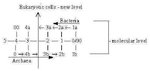 Fig
Ev-add-4 Fig
Ev-add-4 Some oppositions
between the prokaryotic types (Wikipedia):
DNA level:
- Archae: central
DNA have similarity with central
DNA in EKc
- Bacteria: DNA is
more similar to DNA of peripheral
organelles in cytoplasm of EKc.
Invaginated
or immigrated (?) into EKc according
to different
hypothesis. Membranes:
-
Archae special own type with isoprenes
(5C units, with side-chains, thus
of higher complexity). Ether-bonds
(stronger).
- Bacteria: membranes
similar to EKc, fatty acids (linear
chains, 2C units). Ester-bonds (weaker).
Glycerol in membranes:
- Archae:
L-glycerol
- Bacteria: D-glycerol
as in EKc.. Cf. Flagella;
Archae:
central fiber bundles - build from
the base.
Bacteria: hollow tubes
- built from top. The
figure could give the impression
that some kind of neoteny in Archae
contributed to Eukaryotes, a preceding
stage in its "embryology? Levels
of organization from Microcosm to
Macrocosm, just an outline here.
|